Understanding Keratoconus: What It Means for Your Eyes and Vision
Have you ever wondered why your vision suddenly starts to blur or why your glasses no longer seem to work as well as they used to? The answer might lie in a lesser-known, yet significant eye condition called Keratoconus. This progressive eye disease affects the cornea, the clear front surface of your eye, leading to vision changes that can impact your daily life. But what exactly is Keratoconus, and how does it alter the anatomy of your eye?
The Anatomy of the Cornea and Keratoconus
When discussing Keratoconus, it’s crucial to understand the complex anatomy of the cornea and how this condition alters its structure and function. The cornea, a transparent structure at the front of the eye, is vital for focusing light onto the retina, which allows us to see clearly. It’s composed of five distinct layers, each playing a critical role in maintaining corneal integrity and clarity.
The Five Layers of the Cornea
- Epithelium: The outermost layer, acting as a protective barrier against dust, debris, and bacteria.
- Bowman’s Layer: A tough layer that protects the corneal stroma, contributing to the cornea’s strength and shape.
- Stroma: The thickest layer, composed of collagen fibers and keratocytes, responsible for the cornea’s shape and transparency.
- Descemet’s Membrane: A thin but strong layer that protects against injury and infection.
- Endothelium: The innermost layer, maintaining corneal hydration and clarity through fluid regulation.
How Keratoconus Affects Corneal Anatomy
In Keratoconus, the structural integrity of the cornea is compromised. The stroma, which makes up about 90% of the cornea’s thickness and is crucial for its shape and strength, undergoes thinning and weakening. This weakening is thought to be due to an imbalance of enzymatic activity within the cornea and oxidative damage caused by free radicals. As a result, the normal pressure from within the eye causes the weakened cornea to bulge outward.
This bulging leads to a cone-like deformation, primarily affecting the central cornea. It alters the regular curvature of the cornea, leading to irregular astigmatism, where the surface of the cornea is not uniformly curved. This irregularity disrupts the focusing of light onto the retina, leading to distorted and blurred vision.
Molecular and Cellular Changes in Keratoconus
At a cellular level, Keratoconus is characterized by changes in the corneal stromal cells, or keratocytes. There’s a reduction in the number of keratocytes, which impacts the production and maintenance of collagen fibers, crucial for maintaining corneal shape and rigidity. Also, there’s an alteration in the extracellular matrix composition, leading to the weakening of the corneal structure.
The Role of Genetics and Environment
The exact cause of Keratoconus is not fully understood, but it’s believed to be a combination of genetic predisposition and environmental factors. Some research suggests a link to systemic conditions like Down syndrome and connective tissue disorders, indicating a complex interplay of factors contributing to this condition.
Understanding the intricate anatomy of the cornea and the pathophysiological changes in Keratoconus helps in appreciating the complexity of this condition. It underscores the importance of early detection and intervention to manage its progression effectively.
Symptoms and Diagnosis
Keratoconus presents a range of symptoms and requires careful diagnostic processes for accurate identification. Understanding these symptoms and the diagnostic approach can help patients seek timely medical attention.
Detailed Symptoms of Keratoconus
Keratoconus typically develops during adolescence and can progressively worsen. The symptoms vary depending on the stage of the condition:
- Blurred and Distorted Vision: As the cornea becomes more irregular in shape, it causes light rays to focus improperly on the retina, leading to blurred or distorted vision.
- Increased Light Sensitivity: Patients often experience heightened sensitivity to bright lights and glare, which can cause discomfort and visual challenges, especially in well-lit environments.
- Frequent Changes in Eyeglass Prescriptions: Rapid and frequent changes in prescriptions, particularly an increase in astigmatism, can be a sign of progressing Keratoconus.
- Ghost Images: The irregular shape of the cornea can lead to multiple images or ‘ghosting’, often noticed around lights or objects.
- Difficulty with Night Vision: The changes in corneal shape can make it particularly hard to see in low-light conditions.
- Eye Strain and Headaches: Resulting from the constant effort to focus and compensate for the distorted vision.
Diagnostic Approach
Diagnosing Keratoconus involves a combination of patient history, symptom evaluation, and specialized tests:
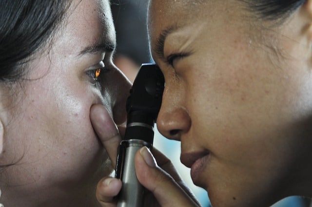
- Comprehensive Eye Examination: An initial step where an optometrist evaluates the overall eye health and vision, looking for signs that suggest Keratoconus.
- Corneal Topography and Tomography: These are advanced imaging techniques that create a detailed map of the cornea’s surface and its thickness. They are crucial in detecting the early stages of Keratoconus and in monitoring its progression.
- Pachymetry: This test measures the thickness of the cornea. Thinning of the cornea is a key indicator of Keratoconus.
- Slit-lamp Examination: Using a specialized microscope, the optometrist can examine the cornea in detail, looking for signs like corneal thinning or a conical shape.
- Retinoscopy: In some cases, a lighted instrument called a retinoscope is used to examine how light is reflected from the retina. This can reveal irregularities in the cornea’s shape.
Early Detection and Regular Monitoring
Early detection and regular monitoring are vital in managing Keratoconus. Progression can be variable; some individuals may experience rapid deterioration, while in others, the condition may progress slowly. Regular follow-ups with an eye care professional help in tracking these changes and adjusting treatment plans accordingly.
Recognizing these symptoms and understanding the diagnostic process empowers patients to seek timely care, which is crucial in managing Keratoconus effectively and preserving vision.
Management and Treatment
Managing and treating Keratoconus involves a multifaceted approach tailored to the severity of the condition and the individual needs of the patient. Understanding the various treatment options can help patients make informed decisions about their eye care.
Non-Surgical Management
- Eyeglasses and Soft Contact Lenses: In the early stages, mild Keratoconus can often be corrected with eyeglasses or soft contact lenses. However, as the condition progresses, these may become less effective.
- Rigid Gas Permeable (RGP) Contact Lenses: RGP lenses are often more effective than soft lenses for moderate Keratoconus. They maintain their shape over the irregular cornea, providing clearer vision by creating a smooth refractive surface.
- Hybrid Contact Lenses: These lenses have a rigid center and a soft peripheral ring, offering the clarity of RGP lenses with the comfort of soft lenses.
- Scleral and Semi-Scleral Lenses: Larger in diameter, these lenses vault over the cornea and rest on the sclera (the white part of the eye), providing comfort and excellent vision correction.
Surgical and Interventional Treatments
As Keratoconus progresses, more advanced treatments may be necessary:
- Corneal Collagen Cross-Linking (CXL): This procedure strengthens the corneal tissue to halt the progression of Keratoconus. It involves the application of riboflavin (vitamin B2) to the cornea, which is then activated with UV light to strengthen the collagen fibers.
- Intacs Inserts or Intracorneal Ring Segments: These are small, curved devices surgically inserted into the cornea to flatten the corneal shape, thereby reducing astigmatism and improving vision.
- Topography-Guided Photorefractive Keratectomy (PRK): In certain cases, PRK is used to reshape the cornea by removing a small amount of corneal tissue, improving the corneal surface and vision.
- Corneal Transplant: In advanced cases where vision cannot be corrected with other treatments, a corneal transplant may be necessary. This involves replacing the damaged cornea with a healthy donor cornea.
Monitoring and Long-Term Care
Regular monitoring is essential for patients with Keratoconus. Follow-up appointments allow for the assessment of disease progression and the effectiveness of treatments. The frequency of these appointments will depend on the severity of the condition and how rapidly it is changing.
Importance of Individualized Treatment Plans
Each patient’s experience with Keratoconus is unique, and thus, treatment plans should be highly personalized. Collaborating closely with an eye care professional ensures that treatments are tailored to meet the specific needs and lifestyle of the individual.
Living with Keratoconus
While Keratoconus can be a challenging condition, with proper management and treatment, most individuals can lead a normal life. Staying informed, adhering to treatment plans, and maintaining regular eye care appointments are key to managing this condition effectively.
At Compton Eye Associates, we are committed to providing our patients with the latest and most effective treatments for Keratoconus. Our team of skilled professionals is here to guide you through every step of your journey towards better eye health.
Your Next Steps
At Compton Eye Associates, we understand the challenges that come with Keratoconus. Our team of experienced optometrists is equipped to provide you with personalized care and the most advanced treatment options. Whether it’s for a comprehensive eye examination, discussing contact lenses options, or exploring treatment for Keratoconus, we are here to support your vision health.
Don’t let Keratoconus blur your vision of the future. Call us today at 800-936-0036 or schedule an appointment with Compton Eye Associates to take the first step in managing your eye health. Remember, early detection and treatment can make all the difference in preserving your vision.
References
- National Eye Institute. (n.d.). Facts About Keratoconus. Retrieved from [https://www.nei.nih.gov/learn-about-eye-health/eye-conditions-and-diseases/keratoconus]
- Mayo Clinic. (2020). Keratoconus. Retrieved from [https://www.mayoclinic.org/diseases-conditions/keratoconus/symptoms-causes/syc-20351352]
- American Academy of Ophthalmology. (2019). What Is Keratoconus?. Retrieved from [https://www.aao.org/eye-health/diseases/what-is-keratoconus]